Its made up of three parts the midbrain pons and medulla oblongata. Anatomy of the Brain What is the central nervous system CNS.
 Duke Neurosciences Lab 1 Surface Anatomy Of The Brain
Intelligence creativity emotion and memory are a few of the many things governed by the brain.

Anatomy of the brain. Anatomy of the Brain. Between the skull and brain is the meninges which consist of three layers of tissue that cover and protect the brain and spinal cord. The brain is an important organ that controls thought memory emotion touch motor skills vision breathing temperature hunger and every process that regulates our body.
The cerebrum is the largest part of the brain. The brain and spinal cord are the two main structures of the central nervous system. The cerebrum which is the largest part of the three main parts making up about 85 of the brain.
The M2-segment is the part in the sylvian fissure and the M3-segment is the cortical segment. It is surrounded by a fluid called cerebrospinal fluid CSF. There are different ways of dividing the brain anatomically into regions.
Ninja NerdsJoin us in this video where we discuss the anatomy of the brain through the use of a model. Join this channel to get access to perkshttpswww. Together the cranium and bones that protect the face are called the skull.
The cranium protects the brain from injury. The human brain is the central organ of the human nervous system and with the spinal cord makes up the central nervous systemThe brain consists of the cerebrum the brainstem and the cerebellumIt controls most of the activities of the body processing integrating and coordinating the information it receives from the sense organs and making decisions as to the instructions sent to the. Underneath lies the brainstem and behind.
It is made up of more than 100 billion nerves that communicate in trillions of connections called synapses. Anatomy of the Brain. It consists of three distinct regions.
The brain has four ventricles connected by cavities and tubes. The brain is one of the largest and most complex organs in the human body. The main functions of CSF are to protect the brain it acts as a shock absorber to carry nutrients to the brain and remove waste from it.
The brain is housed inside the bony covering called the cranium. The two lateral ventricles in the cerebral hemispheres communicate with a third located in the center of the brain. Anatomy of the human brain The largest part of the human brain is the cerebrum which is divided into two hemispheres according to the Mayfield Clinic.
Anatomy of the brain. Although the brain of higher vertebrates undergoes considerable modification during embryonic development these three regions are still discernible. The forebrain midbrain and hindbrain.
Lets use a common method and divide the brain into three main regions based on embryonic development. This amazing organ acts as a control center by receiving interpreting and directing sensory information throughout the body. Its comprised of a left and right hemisphere.
Join us in this video where we discuss the anatomy of the brain through the use of a dissectible model to get a better view at smaller structure. In lower vertebrates the brain is tubular and resembles an early developmental stage of the brain in higher vertebrates. Anatomy of the Brain Overview The human brain is an amazing three-pound organ that controls all functions of the body interprets information from the outside world and embodies the essence of the mind and soul.
The hindbrain the midbrain and the forebrain. There are three major divisions of the brain. The cerebral cortex is what we see when we look at the brain.
Its divided into two halves called hemispheres. The brain is very delicate and is well protected by the skull. Each bump on the surface of the brain is known as a gyrus while each groove is known as a sulcus.
Breathing heart rate body temperature wake and sleep cyclesdigestion sneezing coughing vomiting and 7 swallowing. Horizontal part of the middle cerebral artery which gives rise to the lateral lenticulostriate arteries which supply most of the basal ganglia. The anatomy of the brain is complex due its intricate structure and function.
The two hemispheres are separated by a groove called the interhemispheric fissure. Human Brain Cerebrum touch vision hearing speech reasoning emotions learning fine control movements Cerebellum Co-ordinate muscle Brain Stem relay center movements maintain connecting the cerebrum and cerebellum to posture and balance the spinal cord. The brainstem which is the bodys regulator for all involuntary movement and a relay center for the brain and body.
It is the outermost portion that can be divided into the four lobes of the brain. The CNS consists of the brain and spinal cord.
Lesions can be due to disease trauma or a birth defect. The brain is a very complex organ and even though it is not as complicated as the heart or the lungs it is important to understand the anatomical structure of the brain.
 Ms Brain Lesions Pictures Symptoms And More
Usually medicines can be used to treat the underlying cause.

Lesions on the brain. Usually the lesions of brain are insignificant at first. Brain lesions lesions on the brain refers to any type of abnormal tissue in or on brain tissue. What Diseases Can Cause Lesions on the Brain.
Usually a brain lesion is an incidental finding unrelated to the condition or symptom that led to the imaging test in the first place. A lesion may be localized to one part of the brain or they may be widespread. A lesion is an area of tissue that has been damaged through injury or disease.
Brain lesions are a type of damage to any part of brain. The most common cause of brain lesions is trauma to the brain. On CT or MRI scans brain lesions appear as dark or light spots that dont look like normal brain tissue.
Some examples of brain lesions include abscesses which are caused by infection in the brain and arteriovenous malformations clusters of veins which have grown abnormally. It may be due to trauma or any other disease that can cause inflammation malfunction or destruction of a brain cells or brain tissue. Surgery may be an option in some cases such as when the lesions are caused by a brain tumor.
If the brain lesion is the result of a tumor blocked blood vessel or ruptured aneurysm brain surgery can effectively remove the tumor unclog the. Sometimes lesions and symptoms dont improve even after appropriate diagnosis and proper treatment and the goal is to manage symptoms. At other times the lesions are present in a large part of the brain tissue.
Lesions can also be caused by head injuries strokes multiple sclerosis Alzheimers disease and Creutzfeldt-Jakob disease CJD. Other chemicals and toxins have been associated with brain lesions as well. Injury to the brain can occur either due to fall from height or blowing off some hard object on the head of a person.
A brain lesion is an abnormality seen on a brain-imaging test such as magnetic resonance imaging MRI or a computerized tomography CT scan. Lesions can be due to any disease trauma or it might be the result of some birth defects. Brain lesions can occur due to trauma or a disease process.
That brain damage can affect different aspects of cognition independently and that a locally damaged brain functions identically to a normal brain in its undamaged parts. Surgery to remove the lesion. Sometimes lesions appear in a specific area of the brain.
Lesions on the spine Lesions on the spine are also common in people with MS. While the definition sounds simple understanding. So a brain lesion is an area of injury or disease within the brain.
They may occur spontaneously or develop over a period of time. Brain lesions can arise from trauma or injury benign tumors malignant tumors stroke and other vascular problems infections immune conditions brain cell death or malfunction and ionizing radiation. What diseases can cause lesions in the brain.
Brain lesions can be defined as destruction or damage of any part of brain. Lesions in many brain regions are known to impair both acquisition and retention. At first brain lesions may not produce any symptoms.
Brain lesions may help researchers understanding brain function. This information helps doctors determine the long-term impact of MS. Such lesions might be localized to specific area of brain or might include a large part of brain tissues.
Treatment depends on the type of brain lesion. A brain lesion describes damage or destruction to any part of the brain. Research involving lesions relies on two assumptions.
A T2-weighted MRI scan shows the number of old and new lesions in a specific part of the brain or spinal cord. For example in an extensive and systematic series of studies Thompson and Thorne 1973 trained rats in a visual pattern discrimination task and examined the effects of discrete lesions to numerous brain regions on retention of the learned response. Brain lesions have many different causes that range from traumatic brain injury or disease such as a infection autoimmune diseases abscess stroke genetic disorders or tumors or cancers primary brain cancer and metastatic cancer or brain metastases.
Major types of brain lesions are traumatic infectious malignant benign vascular genetic immune plaques brain cell death or malfunction and ionizing radiation. A brief detail of different conditions that can cause brain lesions is given below. This is because demyelination which is what causes lesions on the nerves is a characteristic sign of MS.
Wait and see if the lesion is not causing problems and is not growing. A brain lesion may involve small to large areas of your brain and the severity of the underlying condition may range from relatively minor to life-threatening. Since the term can refer to any abnormality at all there are numerous causes of brain lesions.
Brain lesions are treated in a number of ways.
The Evolution of the Human Brain. Page 1 of 9 Evolution of Human Brain and Mind The earliest forms of life on earth are about 38 billion years old compared to its 45 billion years age.
 What Is The Evolution Of The Human Brain And Intelligence In Layman Terms Quora
In the 1950s someone with microcephaly might have been a circus attraction but today this condition provides insight into human brain evolution.

Evolution of the human brain. BrainFactsSfN 4 min Your Cursing Cortex. Evolution of human brain mind 1. There are a few major structural differences between human brains and other primates that have contributed to its success in the world.
Pages 179-216 Download PDF. So what makes it special. The expansion during evolution has facilitated the addition of microcircuits leading to the uniqueness of the human brain.
Through evolution human brains have developed into a feat of genetic excellence giving humanity the ability to walk work together and communicate well. Evolution never stops though. The human brain is neither the largest nor most complex brain in the animal kingdom.
This caused the human brain to evolve at a relatively rapid pace. FPGHulton Archive Getty Images One way to determine if brain evolution is in our future is to consider how our brain evolved in the past. The evolutionary history of the human brain shows primarily a gradually bigger brain relative to body size during the evolutionary path from early primates to hominids and finally to Homo sapiens.
As human society became more sophisticated the advantage of a larger brain became more pronounced. The brain has undergone some remarkable changes through its evolution. Origin and Evolution of the Human Mind.
The human brain has been able to reach its current level of complexity and functionality due to evolution. 800000 to 200000 Years Ago. The brain has evolved due to genomics metabolic costs social and ecological drives and linkages from our ancestors the Neanderthals.
Curses swears and cussing are treated in a different way in our brains than our more proper and acceptable speech. Authors Colette Dehay 1 Henry Kennedy 1 2 Affiliations 1 University of Lyon Universite Claude Bernard Lyon 1 Inserm. Because fossilized brain tissue is rare a more reliable approach is to observe anatomical characteristics of the skull that offer insight into brain characteristics.
Researchers at the Howard Hughes Medical Institute concluded that the human brain evolved very rapidly. It will be argued that with the evolution of the human brain we have nearly reached the limits of biological intelligence. A new framework for understanding human brain evolution.
Human brain evolution involves cellular genetic and circuitry changes. Evolution of the Human Brain The human brain can approximately store 125 terabytes of information and can do this through the on average 100000000000 neurons it has and about the 100000000 meters of interconnections of these neurons Cherniak 2012. Over these years in the history of the Earth there was a large climatic shift.
Their research led them to believe that there was considerable selection pressure to evolve the brain into a larger stronger unit. Chapter 8 - Life history changes accompany increased numbers of cortical neurons. Evolution of the human brain Science.
Species that could not adapt to the shifting temperatures and environments quickly went extinct. It is important for an individual to know that due to the stages of human evolution the human brain has become apter at performing higher cognitive functions over time. Genes that control the size and complexity of the brain have undergone much more rapid evolution in humans than in non-human primates or other mammals according to a new study by Howard Hughes.
It led to the modern human brain coming into existence in Africa by about 200000 years ago. This has allowed all human beings to become more complex creatures who are capable of performing a number of highly intelligent procedures. On a genetic level humans have a modified FOXP2 gene which is associated with speech and language development.
The most primitive brains are little more than clusters of cells bunched together at the front of an organism. The human brain in all its staggering complexity is the product of millions of years of evolution. The human variant of the gene SRGAP2 SRGAP2C enables greater dendritic spine density which fosters greater neural connections.
Lateral views of the brains of some mammals to show the evolutionary. The human brain is neither the largest nor most complex brain in the animal kingdom. Homo sapiens of today are not more than 1 00000 years old though hominid line is 3 to 5 million years old.
So what makes it special. 2 The brain initially evolved as a motor program system capable of generating more complex movements then as a temporary memory system able to both play back and produce immediate fear-based reactions to certain situations and finally as a permanent memory system that constructs a model of the world to make predictions about that world. Mammals are 3 to 5 million years old.
They can occur in the cerebral hemispheres and basal ganglia and occasionally in the brainstem and cerebellum Wardlaw et al 2013a. MRI brain diffuse swelling of the brain due to extensive edema of the white matter.

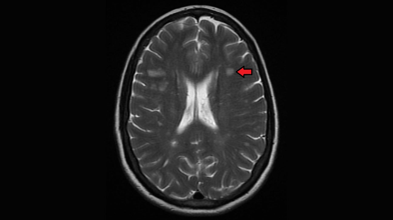
There are several causes of white spots on a brain MRI including small strokes migraines multiple sclerosis MS lupus B12 deficiency a brain tumor such as lymphoma or an infection such as Lyme disease or HIV.

White matter lesions on brain mri. What matter lesions are small groups of dead cells that clump together in the white matter of the brain. This represents as an isolated finding nonspecific white matter disease which in the appropriate patient and with no other characteristic abnormalities on brain MRI would be consistent with MS. White matter lesions on the brain will likely be found on an MRI scan.
When they occur along with other symptoms they are often indicative of a neurological disorder and impaired neurological function. The findings by imaging specialists at NYU Grossman School of Medicine center on small bright spots on scans called white matter hyperintensities. The radiologists report usually further reads that these can be seen in primary demyelinating conditions like multiple sclerosis or in vascular disorders.
White matter signal changes on MRI may also be seen in patients who have infectious and other inflammatory conditions. The presence of these anomalies called white matter hyperintensities characterizes elderly subjects as well as ischemic stroke patients regardless of their age. A new tool for analyzing tissue damage seen on MRI brain scans can detect with more than 70 percent accuracy early signs of cognitive decline new research shows.
The Fazekas scale is used to simply quantify the amount of white matter T2 hyperintense lesions usually attributed to chronic small vessel ischemia although clearly not all such lesions are due to this. They have been reported in the MRI of patients with a history of migraine headaches migraine too is a vascular disorder and that may explain the connection. In many cases CT and MRI imaging studies help pinpoint the location.
It shows brain tissue detail as well as the brain stem and cerebellum posterior brain better than a CT scan. The human brain is made of both gray and white matter. White matter hyperintensities are thought to be caused by small vessel infarcts restriction in blood flow in the.
February 06 2021 An MRI of a human head which can be used to check for white matter lesions. DWI - the posterior limbs of the internal capsules and optic radiations and the central corticospinal tracts within the cerebral hemispheres exhibit diffusion restriction. The structures in the posterior fossa exhibit prominent changes in signal intensity.
White matter lesions are areas of dead cells found within the brain primarily in connective tissue. This classification was proposed by Fazekas et al. A white spot on a brain MRI referred to as white matter or a brain lesion shows an area of injury or disease but can result in a number of diagnoses according to the Mayo Clinic.
Aging is associated with the appearance of increased white spots visualized on brain MRI scans. Information is typically stored and archived in the gray area but the white parts play an important role when it comes to shuttling signals back and forth and retrieving information from one place and bringing it to the next. Introduction White matter hyperintense WMHI lesions are the most common finding in magnetic resonance imaging MRI of the brain in patients with systemic lupus erythematosus SLE.
The potential diagnoses range from harmless and controllable to possibly life threatening. Objective The objective of this article is to determine the clinical factors associated with an increase in WMHI lesion load among SLE patients. Many times I get consulted by patients or their relatives when their MRI brain report reads multiple scattered white matter lesions seen.
Histopathologic lesions were defined. Brain lesions can be caused by injury infection exposure to certain chemicals problems with the immune system and more. Magnetic resonance imaging MRI is a diagnostic test that produces three-dimensional or 3D images of the inside of the body using magnetic fields and computer technology.
This was assessed within subcortical white matter tissue samples harvested from postmortem T 2 magnetic resonance imaging MRI-detected white matter hyperintensities from normal-appearing white matter distant from coexistent MRI-defined hyperintensities and from equivalent areas in MRI normal brains. On CT or MRI scans brain lesions appear as dark or light spots that dont look like normal brain tissue. White-matter lesions are best visible on T2 and fluid-attenuated inversion recovery FLAIR-weighted MRI sequences where they generally appear as symmetric hyperintensities Wardlaw et al 2013b.
A magnetic resonance imaging MRI test which takes pictures of the inside of your brain can show any damage. Changes to white matter will show up super-bright white your doctor may call this. In 1987 1 and at the time of writing late 2016 it remains the most widely used system for describing white matter disease severity in.
Brain lesion on MRI A brain lesion is an abnormality seen on a brain-imaging test such as magnetic resonance imaging MRI or computerized tomography CT.